Comprehensive Guide to Pet Radiography: Enhancing Veterinary Diagnostics
- Written by Business Daily Media

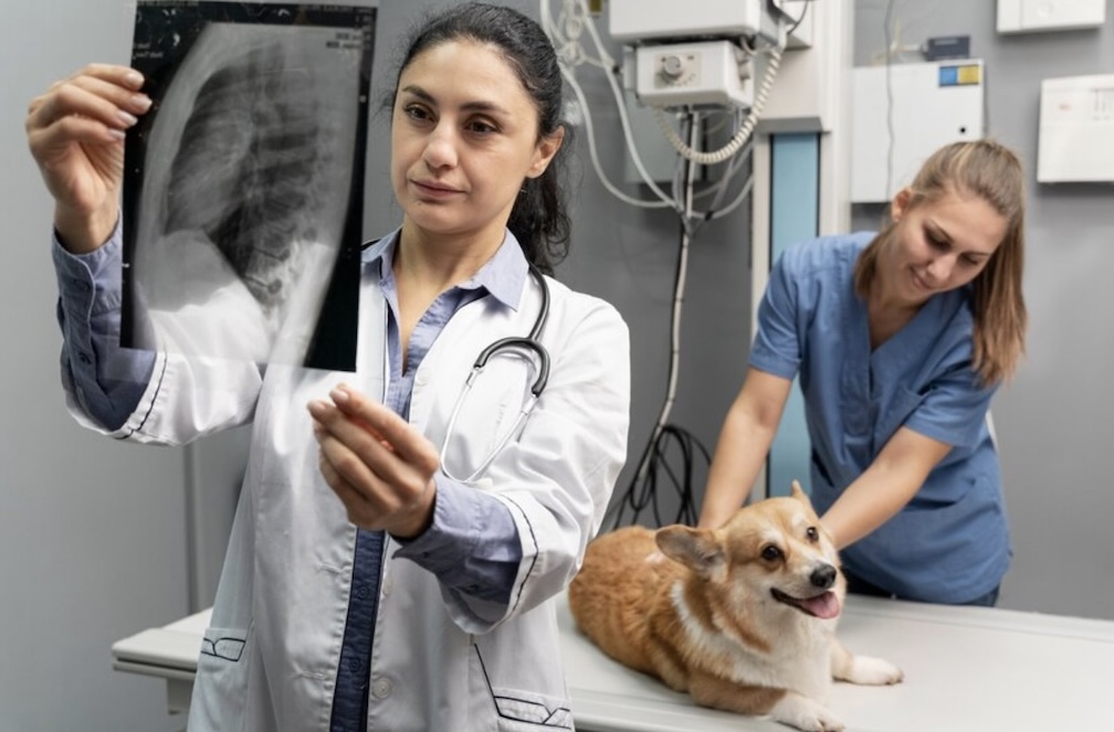
Pet radiography is a non-invasive diagnostic technique that provides detailed images of an animal's anatomy, including bones and organs. It assists veterinarians in diagnosing diseases or injuries in pets effectively and swiftly.
Pet radiography is vital in veterinary diagnostics, enabling a comprehensive internal examination. It identifies diseases or injuries within animals' bodies, enhancing early and precise detection of conditions like bone fractures, tumours, or organ dysfunctions and ensuring effective treatment planning.
Understanding Pet Radiography
History and development of pet radiography
The history and development of pet radiography began in the late 19th century. Today, it's a vital tool in veterinary medicine for diagnosing injuries or illnesses among animals by providing precise internal imaging through pet radiography Sydney.
Basic principles and elements of radiography
Radiography relies on basic principles like differential absorption and geometric distortion. Essential elements include the radiation source, subject, image receptor, processing, and evaluation for creating diagnostic images of the human body's internal structure.
Differences between human and pet radiography
Human and pet radiography both utilise X-rays to visualise internal structures, but they distinctly differ in practices due to anatomical variations, different positioning requirements, and diverse levels of patient cooperation and immobilisation techniques.
Types of Pet Radiography
Traditional Radiography
Traditional radiography is a medical imaging technique that utilises X-ray technology to visualise internal structures. It's often used in diagnosing fractures, detecting infections, examining cavities, or studying anomalies within the human body.
Digital Radiography
Digital radiography is a cutting-edge medical imaging technology. It uses digital X-ray sensors instead of traditional photographic film, improving patients' safety by significantly reducing radiation exposure and quickening the diagnostic process with sharp image capture quality.
Sonography
Sonography, a diagnostic medical procedure, employs the use of high-frequency ultrasound waves to capture dynamic images of the body's internal structures and organs. It assists in tracking diseases and evaluating conditions non-invasively.
Cases Where Pet Radiography is Critical
Diagnosis of bone fractures
The diagnosis of bone fractures typically involves physical examinations and imaging tests like X-rays, CT scans, or MRIs. Symptoms include pain, swelling, and visible deformities. It is crucial to seek immediate medical attention for an accurate diagnosis.
Investigating tumours, infections or diseases
Researchers tirelessly investigate tumours, infections, or diseases using advanced scientific methods. Their goal is to uncover new treatments or cures, enhance understanding of how these conditions affect human health, and improve patient outcomes worldwide.
Examination of heart and lungs
The examination of the heart and lungs involves various procedures like stethoscope auscultation, imaging techniques, and tests to assess their functionality and structure and detect any abnormalities crucial for cardiology or pulmonology diagnosis.
Detection of foreign objects
The detection of foreign objects is essential for public safety. Through systems such as metal detectors and X-ray scanning, unknown or harmful items can be identified and intercepted to prevent potential risks or attacks.
The procedure of Pet Radiography
Description of the radiographic procedure
The radiographic procedure involves exposing a specific body part to a small dose of ionising radiation to produce an image. The radiation highlights internal structures, enabling the efficient diagnosis and treatment of various health conditions.
Preparation process for pets before radiography
Before radiography, pets are usually fasted to clear the digestive system. Sedation or anaesthesia may be administered depending on the pet's anxiety level. Positioning is crucial for accurate images and requires great care and precision.
Risks and considerations in pet radiography
Pet radiography provides essential diagnostic insights but bears certain risks, like radiation exposure. Considerations include animal sedation, handling and positioning challenges, equipment costs, and potential side effects such as injury or stress in animals.
Interpreting Results from Pet Radiography
The basic process of analysing radiographs
Radiograph analysis involves image acquisition, observation for abnormalities, interpretation, and diagnosis. Experts examine contrast variations, identify irregular formations or patterns, compare them with normal radiographic appearances, gather clinical information, and present their conclusions accurately.
Explanation of common findings
Common findings are broadly shared outcomes or results obtained from research or investigations. These could range from medical patterns and scientific phenomena to sociological trends. Such insights typically contribute to a broader understanding and generalizability in various fields.
Importance of follow-up consultation
Follow-up consultations are crucial in healthcare. They ensure effective treatment, monitor progress, manage any side effects, and offer continued support. This undervalued step could mean the difference between recovery and relapse.
Advanced Pet Radiography Techniques
CT Scans for pets
CT Scans for pets are imperative in diagnosing internal injuries or conditions. The procedure, which is non-invasive and painless, provides high-resolution images that aid veterinarians in accurately locating abnormalities within the pet's body for effective treatment strategies.
Ultrasound Imaging
Ultrasound imaging, a non-invasive medical tool, uses high-frequency sound waves to produce real-time images of internal body structures. It aids in diagnosis and treatment monitoring without using harmful radiation or invasive surgical procedures.
Magnetic Resonance Imaging (MRI)
Magnetic resonance imaging (MRI) is a non-invasive medical imaging technique. Using strong magnetic fields and radio waves, MRI generates detailed images of the body for diagnosis or treatment planning purposes without radiation exposure.
Role of Pet Radiography in Veterinary Medicine
Overview of benefits it provides to the veterinary field
Integration of advanced technology in the veterinary field provides numerous benefits, including improved disease diagnosis, efficient treatment methods, enhanced patient management, better training for vet professionals, and overall progress in animal health care.
Current trends and research in pet radiography
Current pet radiography research highlights the growing use of digital imaging technologies and AI advancements, which improve diagnostic precision. Studies also emphasise reducing radiation exposure to ensure both pet and veterinarian safety during tests.
Challenges and likely future developments
Challenges will always be present, driving innovation and adaptation. Likely future developments include advancements in technology, artificial intelligence, renewable energy resources, and stricter environmental regulations confronting climate change at the global level.
Pet radiography FAQs
What is PET radiography?
PET radiography, or positron emission tomography, is a nuclear imaging technique that visualises functional processes in the body. It uses radioactive tracers to detect disease before it shows up on other imaging tests.
What does PET stand for in radiology?
In radiology, PET stands for Positron Emission Tomography. It's a diagnostic imaging technique used to observe metabolic processes in the body, helping to detect diseases like cancer and neurological disorders at an early stage.
Why would a radiologist recommend a PET scan?
A radiologist would recommend a PET scan to evaluate the functioning of organs, identify cancerous activities, ascertain the progress of treatment, and diagnose neurological conditions and heart diseases. It reveals the body's metabolic changes accurately.
What is a PET x-ray?
A PET X-ray, or Positron Emission Tomography scan, is a diagnostic imaging tool that uses radioactive tracers to map out bodily functions like blood flow and oxygen use, offering insights into cell activity and health.


